Article Text

Abstract
Background Diagnostic imaging for low back pain (LBP) without any indication of a serious underlying cause does not improve patient outcomes. However, there is still overuse of imaging, especially at emergency departments (EDs). Although evidence-based guidelines for LBP and radicular pain management exist, a protocol for use at the ED in the Belgian University Hospitals Leuven was not available, resulting in high practice variation. The present paper aims to describe the process from protocol development to the iterative implementation approach and explore how it has influenced practice.
Methods In accordance with a modified ‘knowledge-to-action’ framework, five steps took place within the iterative bottom-up implementation process: (1) identification of the situation that requires the implementation of evidence based recommendations, (2) context analysis, (3) development of an implementation plan, (4) evaluation and (5) sustainability of the implemented practice recommendations. Two potential barriers were identified: the high turnover of attending specialists at the ED and patients’ and general practicioners’ expectations that might overrule the protocol. These were tackled by educational sessions for staff, patient brochures, an information campaign and symposium for general practitioners.
Results The rate of imaging of the lumbar spine decreased from over 25% of patients to 15.0%–16.4% for CT scans and 19.0%–21.8% for X-rays after implementation, but started to fluctuate again after 3 years. After introducing a compulsory e-learning before rotation and catchy posters in the ED staff rooms, rates decreased to 14.0%–14.6% for CT scan use and 12.7–13.5% for X-ray use.
Conclusions Implementation of a new protocol in a tertiary hospital ED with high turn over of rotating trainees is a challenge and requires ongoing efforts to ensure sustainability. Rates of imaging represent an indirect though useful indicator. We have demonstrated that it is possible to implement a protocol that includes demedicalisation in an ED environment and to observe changes in indicator results.
- Emergency department
- Continuous quality improvement
- Evidence-based medicine
Data availability statement
Data are available on reasonable request. The dataset and all study materials (such as the patient information brochures, the poster, the content of the educational sessions and e-learning) are available from the corresponding author on reasonable request.
This is an open access article distributed in accordance with the Creative Commons Attribution Non Commercial (CC BY-NC 4.0) license, which permits others to distribute, remix, adapt, build upon this work non-commercially, and license their derivative works on different terms, provided the original work is properly cited, appropriate credit is given, any changes made indicated, and the use is non-commercial. See: http://creativecommons.org/licenses/by-nc/4.0/.
Statistics from Altmetric.com
WHAT IS ALREADY KNOWN ON THIS TOPIC
Diagnostic imaging for low back pain without any indication of a serious underlying cause does not improve patient outcomes. However, there is still overuse of imaging, especially at emergency departments, and practice variation between healthcare professionals has also been observed.
WHAT THIS STUDY ADDS
This study describes how a knowledge translation framework can help facilitate the development and implementation of an evidence-based protocol into practice.
HOW THIS STUDY MIGHT AFFECT RESEARCH, PRACTICE OR POLICY
The iterative implementation approach, applied in this study, might inform others to tackle practice change at their emergency department. This study also provides other researchers and clinicians with imaging data for benchmarking, as appropriate rates of justifiable CT scans and X-ray images at emergency departments are lacking in the current literature.
Background
Low back pain (LBP) is the number one cause of years lived with disability worldwide, with a point prevalence of 9.6% and an incidence rate of 3951 new cases/100 000 individuals per year.1 2 In Belgium, the situation seems even worse with a point-prevalence of 18.2%.1 In approximately 10% of the cases, the pain and functional impairment will continue beyond 3 months after onset, which results in high healthcare costs and work absence.3 4 Therefore, appropriate management of acute LBP and investment in the avoidance of chronicity is crucial.
In the large majority of patients with acute onset LBP and radicular pain, pain and function are likely to recover spontaneously within weeks. Most often, acute LBP is not attributable to a specific cause, but rather represents an acute mechanical overload leading to symptoms because of reduced load bearing capacity.5 Reassurance and activation are most accurate in those situations and referral to specialist medical care is not required on the condition that a competent healthcare professional has ruled out potentially worrisome pathologies that might potentially endanger the patient when missed (also called ‘red flags’).6 In the absence of red flags, imaging does not affect management.7 8 On the contrary, it has been shown that referral of these patients for diagnostic imaging or for a medical specialist consult may rather facilitate chronicity, disability and overmedicalisation.9 10 Imaging, however, is still being overused in LBP.11–14 Evidence indicates that diagnostic imaging for LBP without any indication of a serious underlying cause does not improve patient outcomes and can in fact be harmful to patients due to radiation exposure on the one hand and imposing a sickness label on patients on the other hand.15 16
Patient factors, such as fears and beliefs, often contribute to LBP patients visiting an emergency department (ED), whether or not they had prior contact with and adequate advice from their general practitioner (GP). Patients with red flag conditions and radicular pain with significant weakness should indeed be referred for assessment in an ED aiming at swift diagnosis and concordant therapy to avoid harm. On the contrary, patients with non-red flag acute LBP and non-alarming acute radicular pain par excellence should be managed in primary care. However, they still often go to the ED,17–19 where the use of undue imaging continues to occur at high rates.20
At the ED of the University Hospitals Leuven, a large tertiary centre in Belgium, the care for patients with LBP and radicular pain was initially unstructured. This resulted in considerable practice variation between attending physicians and subsequent confusion among nurses. Clinical practice guidelines are particularly helpful in addressing practice variation because they aim to describe appropriate care based on the best available scientific evidence and broad consensus while promoting efficient use of resources.21 Despite the existence of international evidence-based clinical practice guidelines for the management of LBP and radicular pain, a locally accepted protocol or guideline was not available until 2012. In 2012, after identifying this as a problem, a multidisciplinary team of specialists developed such a protocol aiming to improve patient care. This included better screening for alarming situations while reducing unnecessary specialist actions, such as diagnostic imaging and specialist clinic appointments in non-alarming situations. The protocol also emphasised patient education and activation and ensured sufficient comfort. As this protocol represented a significant change in practice for a large group of healthcare professionals rotating in the ED, a coherent set of strategies was required to ensure effective implementation of the protocol in practice. The current paper aims to describe the process from protocol development to the iterative implementation approach and explore how it has influenced practice.
Methods
Implementation scientists have argued that the use of frameworks help to increase the likelihood of successful implementation and to reduce the evidence-practice gap.22 23 While there is increasing interest in using frameworks, few implementation projects have made good use of these.22 24–26 Researchers have concerns about the challenges of selecting the most suitable framework for a given project within its local context.27 For the current project, the authors chose the well-known ‘knowledge-to-action’ (KTA) framework,28 29 that describes a process for implementing evidence into practice, also called a ‘process model’.28 30 This KTA framework was adapted to meet the local needs of health professionals working in guideline implementation within the Belgian setting.31 The following five steps were taken: identification, context analysis, development of action plan, evaluation and sustainability. Next, tasks were embedded for each of the five steps (see figure 1). The rationale behind these steps and detailed descriptions of the adaptation process are published elsewhere.31 All tasks were based on the KTA framework, except for the following tasks which were added based on an exploratory literature search and the implementation facilitators’ professional experiences: ‘select guideline recommendations’ (in step 1), ‘screen existing initiatives’ (in step 2) and ‘integrate new practice in routine care’ (in step 5). Further, additional details were provided for steps deemed too abstract or general by the implementation facilitators and practical hands-on tools were included to facilitate some of the steps. All steps are described below, and more details can be found in Peters et al.31 The SQUIRE 2.0 was used to standardise the reporting of this project32 (completed checklist in online supplemental appendix 1).
Supplemental material

A five-step approach for guideline implementation.31
Step 1: identification
A Multidisciplinary Spine Working Group (MSWG), consisting of two physical medicine and rehabilitation specialists, a neurosurgeon, an orthopaedic surgeon, a physiotherapist and a care programme delivery manager, was established to analyse the ‘evidence-practice’ or ‘know-do’ gap and to develop clinical recommendations. This know-do gap was initially identified via an audit of the electronic medical record system, which showed that the majority of patients received care that was not in accordance with evidence-based guidelines and that management of LBP at the ED was subject to huge practice variation. This led to several discussions within the MSWG, who tried to find reasons behind the lack of evidence-based practice and high practice variation. The MSWG concluded that part of the variation was explained because many patients were initially assessed by the attending trauma surgeon, trained not to miss post-traumatic injuries. Hence, in most patients with LBP, X-ray imaging of the lumbar spine was ordered. Likewise, all patients with radicular pain underwent CT scans of the lumbar spine. Further management then depended on which specialty was consulted to take over. Patients with radicular pain would wait for hours for the neurosurgery trainee on call, who then made long-term appointments for additional MRI and a surgery clinic visit, disregarding the potential for spontaneous recovery. There was no strategy for conservative management or advice, nor was there any focus on adequate patient counselling, and concepts of reassurance and maintaining activity were simply not conveyed. Finally, red flags were not systematically investigated.
In order to reduce practice variation and include evidence in patient management, the MSWG designed a protocol based on evidence-based guidelines33–38 and in keeping with organisational aspects of the ED and the hospital. The protocol is available on request. Briefly, it includes a structured intake of patients by the attending emergency physician or internal medicine specialist (instead of the trauma surgeon, as this would give the wrong message of a structural problem to be solved), who will subsequently (1) triage the patient via consecutive steps (screen for red flags, rule out or address radicular pain and/or appropriately address axial pain), (2) when alarming situations are ruled out, ensure reassurance by accurate information, provide sufficient pharmacological pain control and emphasise the maintenance of activity whenever possible and (3) when alarming situations are ruled out, refer acute low back related problems to the primary care setting where they can be perfectly managed, including adequate information for the GP. In addition, the protocol includes the establishment of an accessible hotline for GPs to timely refer patients entering the subacute stage and that seem to be in need for specialist care to avoid chronicity. Finally, a joint neurosurgery/orthopaedic surgery on-call system was set up to address patients with red flags and/or radicular pain with significant weakness.
Step 2: context analysis
Within the MSWG, barriers and facilitators for implementing the protocol were identified via group discussions. Two modifiable barriers to implementing the newly-developed protocol were perceived as priority barriers to address.
First, the University Hospitals of Leuven ED worked as an open system in which several specialists consult a patient, with one specialty being assigned responsibility for the patient. Physicians are predominantly trainees in rotation, who frequently change departments and hospitals, potentially bringing in suboptimal habits from other hospitals regarding patient management. At the moment of the protocol’s introduction, no other hospital in Belgium used an evidence-based protocol for this population.
Second, healthcare professionals at the ED seemed to be inclined to order diagnostic imaging to meet patients’ and GPs’ expectations. They believed that if they did not offer imaging to patients, their GPs would refer them to a different hospital, or the patient would go elsewhere to get imaging done. Also, healthcare professionals felt that they had to plan investigations and clinic appointments to offer the patient something tangible. Thereby, they disregarded that offering imaging and follow-up appointments are associated with a wrong message to the patient and contradicts the philosophy of spine specialists at the spine clinics.
Step 3: development of an implementation plan
An implementation plan, including multiple strategies, was developed based on literature on existing implementation strategies,39–42 local barriers and context, and was discussed in a group of relevant stakeholders. In order to tackle the first barrier on task division and practice variation, educational sessions were perceived to be most useful. In these sessions, the most important items and their rationale and underlying evidence were explained to physicians and ED nurses: accurate triage without need for imaging, unequivocal guidance on imaging and calling the spine care team in case of alarming situations and guidance on reassurance, comfort management, activation and referral to primary care in non-alarming acute situations. The new protocol was made easily accessible on the hospital’s intranet under the spine care programme pages and this was broadly communicated. The second barrier (patients’ and GPs’ expectations on imaging) was addressed by producing patient information brochures and making template back-referral letters for GPs available, by a explanatory letter mailed to GPs and by organising a symposium for GPs. The MSWG realised that they had to target all stakeholders, including patients and GPs. All feedback from associated healthcare professionals and patients in letters and conversations was documented to the maximum extent and discussed in the MSWG meetings. A summary report was made of the discussions that took place at the GP symposium. GPs at the symposium replied that they very much welcomed this initiative and had been waiting for it. They felt that the old habits were counteracting their advices to patients and that the new protocol, therefore, strengthened their approach. All implementation strategies are outlined in box 1.
Implementation strategies 2012–2018
Educational sessions
.
Description of intervention: Based on the protocol, rationale and aim, the content of educational sessions was developed by the Multidisciplinary Spine Working Group. The MSWG organised several face-to-face interactive sessions for all disciplines involved in managing the target patient population at the emergency department (ED). This took place in 2012, 2013 and 2014 and was reboosted in 2018.
Target audience and number of participants: Physicians and trainees of emergency medicine and internal medicine, orthopaedic surgery, neurosurgery, physical and rehabilitation medicine, trauma surgery (total: 26 physicians, >100 trainees) as well as ED nurses (>100).
Patient information brochures
Description of intervention: Brochures contain key messages of importance to patients: how potentially treatening situations are ruled out, how to obtain comfort, why staying active is important, why imaging is not always relevant and how follow-up is organised with the general practitioner. A separate brochure was created for low back pain and non-alarming radicular pain. The brochures were made available at the ED for handing out to the patient by the attending physician. Brochures were also made available on the spine care program’s webpage.
Target audience and number of participants: Patients with non-alarming low back and non-alarming radicular pain at the ED, approximately 350 patients/year.
Symposium for GPs
Description of intervention: On 24 November 2012, the new protocol and its rationale were presented in a symposium for GPs.
Target audience and number of participants: All GPs from the hospital’s hinterland were invited. Approximately 100 GPs attended the symposium.
Letters for GPs
Description of intervention: The new modus operandi and its rationale was briefly explained in a letter, that also contained the reference of the webpage at which more information could be found.
Target audience and number of participants: the letter was sent to all GPs in the hospital’s hinterland.
Step 4: evaluation
From supervising in the ED, the surgeons that were part of the MSWG knew that imaging was clearly often being prescribed in situations that did not require any imaging according to evidence-based guidelines. This manifested itself particularly in patients with non-alarming acute LBP that would get a lumbar X-ray and patients with acute radicular pain that would almost routinely receive a CT scan of the lumbar spine. It was therefore expected that bringing practice in accordance with guidelines would lead to reduced imaging in these cases. As imaging orders are easily retrievable, the rate of X-ray and CT scan orders in patients with a low back related presentation was an easy target for developing an automated though relative indicator of protocol adherence, whereas we could not think of other indicators that were eligible for automated calculation based on registered data. Through the spine surgery consults that were requested by the emergency and internal medicine physicians in case of doubt or in case of a presumed alarming sign, we could easily observe how high rate of imaging orders and spine surgery consults were associated with lower adherence to the protocol and vice versa.
Three months after the introduction in February 2013, two MSWG members checked the records of a list of relevant patients that came to the ED with LBP and/or radicular pain in the preceding months. They found that the protocol had been accurately applied in the majority of cases and imaging was justified when it had been ordered. In the minority of cases that deviated from the protocol, the trainees and their supervisors were contacted and the new protocol was explained once more. The result of this check was considered positive. A similar check was repeated in 2014, with similar results. To monitor the implementation success continuously, indicators were developed and programmed that reflect the number of X-rays and the number of CT scans of the lumbar spine in the numerator and the number of patients entering the ED for a lumbar spine-related problem in the denominator. In order to enable the calculation of the denominator, free text spine-related diagnoses entered at discharge from the ED had to be structured into detectable categories. Both indicators were included in an automated feedback report, containing also other spine care program-related indicators outside the ED, which was sent to the MSWG on a monthly basis as from October 2015. Results were regularly discussed in MSWG meetings and fed back to the emergency and internal medicine departments.
Step 5: sustainability
Approximately 2 years after the implementation, the use of CT scans started to fluctuate and the decrease of X-rays tempered. Also, surgery trainees on call for spine reported more frequently that they were called by their emergency and internal medicine colleagues upfront. The turnover of high numbers of trainees involved in seeing patients in the ED, applying habits they had picked up elsewhere, was considered responsible for this. However, the 3-month rotation schemes made it impractical to organise training sessions with adequate frequency. Therefore, the action plan was adjusted (see box 2). A compulsory e-learning replaced the face-to-face educational sessions and a eye-catching poster was designed to hang up at critical places at the ED to make the key messages of the protocol constantly visible to the staff. Moreover, an update of the patient information brochures was deemed necessary to make them a bit more appealing for patients.
Implementation strategies in 2018–2019
E-learning
Description of intervention: The protocol’s essential content and rationale were briefly explained in a 15 min e-learning. The hospital e-learning system allows making the e-learning a prerequisite before starting the rotation.
Target audience and number of participants: All emergency and internal medicine trainees need to have followed the e-learning before an emergency department (ED) rotation. All neurosurgery and orthopaedic surgery trainees need to have fulfilled the e-learning at the start of their residency.
Poster
Description of intervention: The essential messages of adequately triaging patients, not performing imaging in non-alarming situations and correctly counselling patients were included in a catchy poster hung up in the ED.
Target audience and number of participants: Trainees in emergency and internal medicine rotating in the ED.
Update of patient information brochures (which were developed in 2012)
Results
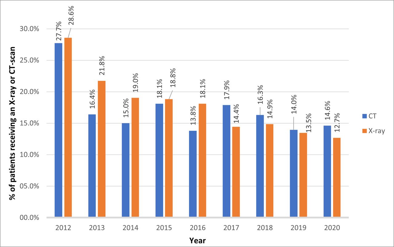
Figure 2 shows the performance of both indicators over the time period of 2012–2020. The introduction of the protocol led to a decrease in imaging orders, after which it fluctuated due to constantly new trainees coming on the ED. Of note, there are no benchmark data available in literature on the rate of justifiable CT scans and X-ray images of the lumbare spine when guidelines are being followed in an ED. It is expected that in a certain percentage of patients imaging is warranted because of suspicious non-degenerative underlying pathology. We observed a significant decrease of both percentages from over 25% before implementation of the new protocol (February 2013) to 13.7%–18.1% for CT scan use and 12.7%–21.8% for X-ray use. Variability is secondary to a certain heterogeneity of the population entering the ED, but also to variable adherence to the protocol.
Percentage of patients admitted at the emergency department with back problems receiving an X-ray or CT scan of the lumbar spine at the University Hospitals Leuven.
The reduction in imaging use at ED was discussed beforehand with the radiology department, who saw no problem and welcomed the initiative. Feedback from the team (trainees, nurses and physicians at ED and GPs) was largely positive (though the feedback was not collected in a systematic and structured manner). There were no costs associated with the implementation, rather it was associated with a capacity gain. Although we did not get feedback from all GPs—and hence, might have lost some referrals—feedback from most GPs was very positive and we did not see a decrease in patient numbers.
After the introduction of the compulsory e-learning and the poster at the ED, implemented at the end of 2018, a subsequent decrease in imaging use was observed, to 14.0%–14.6% for CT scan use and 12.7%–13.5% for X-ray use.
Discussion
The present paper describes the implementation of an evidence-based protocol for managing patients presenting with LBP and lumbar radicular pain at the ED of a large tertiary hospital in Belgium. The process impacted a large group of patients as well as healthcare professionals inside and outside the hospital, and therefore, constituted an important change. After several years, the change is largely accepted and normalised into the hospital, and informal feedback from stakeholders remains positive. Given the latter is fragmented and potentially biased, ongoing evaluation is crucial. Limiting the use of imaging to situations where it may have an impact on medical management is a key element of the new protocol, and we, therefore, chose to monitor the implementation success by the rate of X-rays and CT scans of the lumbar spine ordered for these patients. Meanwhile, the monitoring output has led to additional actions and, hence, is considered useful. At present, management of LBP and radicular pain at the ED can be considered in accordance with international guidelines, and other hospitals from within the hospital network are following our example.
The huge burden caused by LBP worldwide1 and its growing magnitude alongside the increasing and ageing population urged the Lancet to issue a call for action in 20182 . LBP-related problems are not only associated with a huge prevalence, but also with a poor relation between imaging findings and pain generators43 and an overemphasis on medical diagnostics and interventional therapies often leading to poor results.44 At present, the biopsychosocial model introduced by Waddell45 is gaining importance, and the impact of psychosocial risk factors has been confirmed in several studies.46 47 Current international guidelines advocate the importance of accurate triage and, in case of non-alarming situations, demedicalisation by reassurance, comfort measures and activation.33–38 48 The implementation of these guidelines,representing current best evidence, in primary care, hospital care as well as EDs probably constitute the best and maybe singular action to tackle the Lancet’s call. Since EDs often act as a bridge between primary and hospital care and given the often acute presentation of low back related problems, the importance of correct actions and messages to patients at EDs cannot be underestimated. Guidelines and care pathways are excellent means to introduce common vocabulary, reduce practice variation, optimise use of resources and hence, improve quality of care. In 2017, the Belgian Healthcare Knowledge Center issued a care pathway for the management of LBP and radicular pain that can be consulted at www.lowbackpain.kce.be and that is in line with the protocol that was introduced earlier in our ED.49
Identifying and managing barriers to change is an important element of change management. When the protocol was in its development phase, a few older colleagues warned us that patients would not accept receiving no imaging and that GPs would stop referring their patients to our hospital. As it appeared in the conversations with GPs at the symposium, this belief was a serious underestimation of the adherence to new evidence by the primary care field, and the GPs told us they had been waiting for our initiative and we were the ones that had been counteractive in their attempts to educate patients. This illustrates that it is important to include all stakeholders in such a change process. A recent literature review also showed that stakeholder engagement is increasingly used in guideline implementation.50 After implementation of the protocol we never observed a drop in LBP patients coming to the ED. A more difficult barrier to tackle was the high turnover of trainees rotating at the ED and involved in managing the target patient population. Since bedside teaching plays a huge role in medical training, trainees would continue to bring in habits they learnt from external specialists, that at the time had not invested in adhering to guidelines for LBP management in their hospital. The compulsory e-learning before trainees could start their ED rotation meant an enormous help. We also learnt from this that it was essential to include the nursing staff at the ED. They play a moderating role towards the trainees, they provide continuity in the care and are essential in organising unambiguous communication with the patients.
The current paper describes the implementation in accordance with a modified ‘KTA’ framework, with five steps in an iterative bottom-up process. At the start, consulting of evidence mainly focused on getting the protocol right, and the inclusion of an implementation scientist followed later. This illustrates that the essential implementation steps outlined in the methodology described by Peters et al.31 are intuitive and robust. At the same time, although the implementation is considered sufficiently successful, it took us several years. The process might have happened more efficiently if the implementation steps and intermediate goals had been clearly defined upfront. In particular, the acting on the evaluation results to enhance sustainability was initially insufficient and resulted in a temporarily poorer performance of the imaging indicator. In this context, it helped that as from 2017 with the advent of the guideline and pathway issued by the Belgian Healthcare Knowledge Centre, complementary implementation strategies in the primary care setting were being initiated, raising attention again to our own project. Also, the efforts to obtain Joint Commission International accreditation for the hospital spine care programme in 2019 boosted the normalisation into standard care of all spine-related protocols among all collaborators in the entire hospital.
Of note, the programme has never intended the CT scan and X-ray indicators to drop to 0%. In this regard, the indicator is indirect and not ideal. Imaging will always be required in patients with red flags and patients with radicular pain presenting with significant motor weakness. The expected rate of justified imaging orders depends on the hospital context in terms of referral patterns and patient attitudes. There are no literature data available on this, and we learnt that the rates in our ED could safely drop to 12%–13% for X-rays and to 13%–14% for CT scans of the lumbar spine. However, when patients with non-alarming problems would be triaged and stay in primary care, which could be considered the ultimate situation if the educational goals of our program succeed, the imaging indicator values would increase again. Therefore, the value of the indicators is in their trends rather than in the values themselves.
Until recently, there were no comparative studies looking into LBP management at EDs. However, in April 2022 a study got published and the authors found a rate of 11% X-ray use and 0.5% CT scan use. Yet, this was after exclusion of red flag patients.51 Future research could explore the use of an appropriateness score when ordering imaging for LBP in EDs52 and explore how general practice53 and ED can work together in their struggle to reduce imaging.
This is a description of a process that spanned almost ten years. While this illustrates that implementation is an ongoing process rather than one event, we do realise that some variables could have been registered more accurately. The manual screening of patient records took place twice, but was not formally documented. Feedback from patients, nurses, trainees and consultants was based on informal conversations, and could have been organised in a more structured way. Eventual outcome data are not available. We have no means to screen for patients that did go to another hospital after feeling insufficiently helped in our ED, and red flags may have been missed without us being able to capture this. We are aware of one patient with a red flag condition that was subsequently detected in one of our spine clinics and emphasise in our program that red flags should be screened for at every contact. We are not aware of any patient being harmed by implementing this protocol. On the contrary, red flags are being screened for more effectively and patients get better education on the nature of their problem in non-alarming situations.
One additional indicator could have been the mapping of further appointments post-discharge, that should have marked a shift from hospital follow-up to primary care follow-up. Measurement of changes in patient behaviour and beliefs or back pain outcomes would have illustrated the ultimate goal, but were unrealistic and beyond the means associated with this project. One main question is whether there could be alternative explanations for the indicator change we measured in terms of spontaneously changing patient profiles. However, the reduction in imaging orders was quite brisk and could not have been explained by natural inflow changes. Also, the implementation meant quite a revolution at the time, and was the ‘talk of town’ for involved healthcare professionals. Although the latter is not explicitly documented, we know that the change was real. The current paper provides colleagues with an illustrated case to trigger them towards a similar guideline adherent protocol and at the same time provide them with the imaging data for benchmarking.
Conclusion
The implementation of a new evidence-based protocol for the management of LBP and radicular pain in a busy tertiary hospital ED with a high turn over of rotating trainees is a challenge and requires monitoring and ongoing efforts to ensure sustainability. Rates of imaging represent an indirect though useful indicator. We have demonstrated that it is possible to implement a protocol that includes demedicalisation in an ED environment and to observe an influence on practice.
Data availability statement
Data are available on reasonable request. The dataset and all study materials (such as the patient information brochures, the poster, the content of the educational sessions and e-learning) are available from the corresponding author on reasonable request.
Ethics statements
Patient consent for publication
Acknowledgments
The authors would like to thank all other stakeholders involved in this initiative.
References
Supplementary materials
Supplementary Data
This web only file has been produced by the BMJ Publishing Group from an electronic file supplied by the author(s) and has not been edited for content.
Footnotes
SP and KJ are joint first authors.
Contributors SP, KJ, PVW, SR, SS, LM, JD, AS, A-IVdB, JV and BD were involved in the study design, data collection and analysis. SP, KJ and BD wrote the first draft of the manuscript and all authors approved the final version of the manuscript. BD acts as guarantor for this study.
Funding The authors have not declared a specific grant for this research from any funding agency in the public, commercial or not-for-profit sectors.
Competing interests None declared.
Patient and public involvement Patients and/or the public were not involved in the design, or conduct, or reporting, or dissemination plans of this research.
Provenance and peer review Not commissioned; externally peer reviewed.
Supplemental material This content has been supplied by the author(s). It has not been vetted by BMJ Publishing Group Limited (BMJ) and may not have been peer-reviewed. Any opinions or recommendations discussed are solely those of the author(s) and are not endorsed by BMJ. BMJ disclaims all liability and responsibility arising from any reliance placed on the content. Where the content includes any translated material, BMJ does not warrant the accuracy and reliability of the translations (including but not limited to local regulations, clinical guidelines, terminology, drug names and drug dosages), and is not responsible for any error and/or omissions arising from translation and adaptation or otherwise.