Article Text

Statistics from Altmetric.com
Introduction
Emergency airway management is a critical life-saving procedure required for patients in various acute conditions. Airway management outside the operating theatre is associated with serious consequences.1 Common complications of airway management include hypoxaemia, dysrhythmias, cardiovascular collapse, oesophageal intubation, aspiration and death.2
The 4th National Audit Project of the Royal College of Anaesthetists found that more than 30% of emergency department (ED) patients and 60% of intensive care unit patients with an airway related incident suffered death or brain damage, compared with only 14% of patients in controlled settings of an operation theatre.2 3
In the ED, rapid sequence intubation (RSI) is the most commonly used technique of intubation. It involves the administration of an induction agent immediately followed by a paralytic agent.
The patients requiring intubation in the ED are critically ill. They may be hypotensive, hypoxaemic or may respond differently with drugs used for intubation. The incidence of complications in critically ill patients requiring airway management is 40%.4 Hypoxia5 (25%) is the most common complication in these patients followed by hypotension (10%–25%).5 6 Cardiac arrest occurs in about 1.5%.7 Hypoxia due to oxygen desaturation can lead to arrhythmias, hypotension, hypoxic brain damage and cardiac arrest particularly when the saturation falls below 70%.7–10 More than one attempt, preintubation saturation of <93%, and RSI duration more than 3 min are associated with increased incidence of hypoxia.11 Hence, time to intubation is a crucial quality indicator and reference standard for assessing emergency airway management.
Our institute is a tertiary care referral centre and the ED caters to about 450 patients per day. Approximately 10 intubations are performed every week in the medical emergency and RSI technique is used in most of the cases. Our institute runs an academic emergency medicine residency programme. RSI in the ED are performed by emergency medicine junior resident physician under the direct supervision of a senior resident physician. Failed intubation is defined as three unsuccessful laryngoscopy and laryngeal mask airway (LMA) would be inserted or surgical airway secured. As most of the intubations are done by academic residents, we felt the need to improve emergency airway management in the ED and focus on reducing the intubation time (the time from administration of paralytic agent to successful endotracheal (ET) tube insertion) using the quality improvement (QI) methodology.
Methods
A multidisciplinary QI team consisting of a mentor, an academic consultant, a senior resident physician, nurse educators and nursing officers was formed to decrease the intubation time in RSI. We used the Point Of Care Quality Improvement methodology developed by the WHO of South East Asian Regional Office.12 We collected baseline data from 15 patients who underwent RSI by calculating the time between the paralytic agent administration and successful ET insertion. The median time was found to be 300 s (IQR 210 s).
The aim of the QI project was to decrease the median time of intubation from administration of paralytic agent to the successful passage of ET tube into the trachea by 40% from baseline 300 s in patients undergoing RSI in the ED, within 3 months.
The team used process mapping and fish bone analysis to identify the bottlenecks of delay in intubation. The change ideas were tested after discussion with the team members. Three Plan-Do-Study-Act (PDSA) cycles were tested and successfully implemented. In each meeting, small achievements were celebrated and team members were encouraged by the mentor and consultant. No patient was involved in the design, conduct, or reporting of this study.
Measurement
The data were collected by the nursing officers of the QI team when they were on duty but not directly involved with the care of the patient undergoing RSI. Round the clock shift duties made it extremely challenging to collect data of all the intubations performed in the ED. Resident roster is entirely different from the nursing officers. This ensured that a different performer was assessed each time.
The nursing officer would switch on the stop watch on administration of the paralytic agent. The end point was successful tracheal intubation—the time when End Tidal CO2 (ETCO2) reading was obtained or at the end of five point auscultation, if ETCO2 was not available.
Data analysis
We plotted the time interval on run chart for analysis. When a shift was identified on the run chart, the median was recalculated. A shift is defined as ≥6 consecutive data points on the same side of the median.13 We calculated the baseline median for the initial 15 patients from 19 December 2018 to 31 January 2019.
Understanding and analysing processes of care
RSI is the most commonly used technique in the ED. The steps of RSI include: preparation, preoxygenation, pretreatment, administration of induction and paralytic agent, positioning of the patient, laryngoscopy and placement of the tube and postintubation care.14 We performed process mapping from the decision to perform RSI till successful ET intubation. The process mapping was drawn and bottle necks identified in a team meeting (figure 1). Our interest was to reduce the duration from step 4 to step 6 that is, from paralysis to successful placement of ET tube.
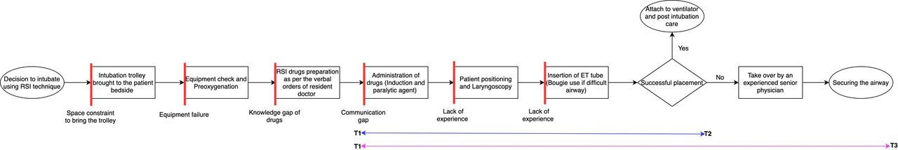
Process flow diagram with bottlenecks highlighted in vertical red lines. T1 as denoted was the start time of measuring and T2 the end time in successful first pass intubation. In case of reattempt, the end point considered was T3. Change ideas were planned as per the problems highlighted in the flow diagram. ET tube, endotracheal tube; RSI, rapid sequence intubation.
After the resident physician decides RSI has to be performed on a patient the nursing officer is informed. The intubation trolley is brought to the patient bedside and preoxygenation is started. Equipment required for RSI are checked. Pretreatment is done depending on the patient’s profile. Induction agent and paralytic required are prepared and administered by the nursing staff on verbal order of the resident physician. The patient is positioned and laryngoscopy is performed about 60 s after administration of the paralytic agent (succinylcholine: 45 s and rocuronium: 60 s). ET tube is inserted and the position is confirmed by either ETCO2 detector or five-point auscultation technique depending on the availability. Bougie is inserted into the trachea in case of difficult intubation and ET tube is railroaded on it. Once the tube position is confirmed, the ventilator is connected and postintubation care is started. If the ET is not positioned in the trachea, further attempts are made by an experienced senior resident. If three attempts fail, laryngeal mask is inserted or a surgical airway (cricothyroidotomy) is performed at the front of the neck. Patients who required LMA or surgical airway were excluded from the project.
The major bottlenecks identified were lack of formal training of resident physicians and nursing staff and communication gap between the resident and nursing staff. We used fish bone analysis to identify the causes of delay in terms of people, place, process and policy(table 1).
Fish bone analysis
Strategies
After several discussions with the team members, we came up with change ideas based on the process mapping and fish bone analysis. PDSA cycles were used to test the change ideas.
PDSA 1: training of frontline residents and nursing officers (class room training)
In the first change idea junior residents and nursing officers of one shift underwent a simulation-based teaching on RSI in the seminar room of the ED. The emergency medicine senior resident and faculty from college of nursing conducted the training session. The frontline team was sensitised about the importance of reducing oxygen desaturation time. Their skills in laryngoscopy and intubation were improved by practising on a manikin under direct supervision of the senior resident and faculty. Each resident was given individual feedback. Through a simulated session, the nursing officers were taught how to facilitate the airway management ensuring all drugs and equipment are ready and functional. Data were collected over 4 weeks and analysed.
PDSA 2: sensitisation of critical care area residents and nursing officers (on-site training)
In order to ensure practice change, short (lasting 10–15 min) on-site training and awareness sessions were organised by senior residents and nursing officers’ mentor for on duty residents and nursing officers working in critical care areas of the department. About 10–12 healthcare workers were part of each on-site training. Steps of intubation, logistical issues, drug and equipment checklist were discussed and local issues were resolved. Three such sessions were organised to cover different shift teams. We ensured that data were collected only after the on-site training of healthcare workers.
PDSA 3: streamlining processes by introducing airway drug checklist and ensuring availability of difficult airway equipment
Inputs from the nursing officers were taken during the on-site training and accordingly an RSI drug checklist and bougie was introduced in the resuscitation trolley. The RSI drug checklist contained the doses of frequently used drugs during intubation. The drug list was pasted on the lid of the drug tray ensuring easy check by the nursing team. This prevented medication dosing error and delay between the administration of induction and paralytic agent. Bougie or ET airway introducer useful in the management of difficult airway was not a part of the resuscitation trolley routinely. Making bougie a compulsory part of resuscitation trolley made sure intubation was quicker even in cases of difficult airway.
Results
After the implementation of change ideas, there was a significant reduction in the time to intubation (table 2). At the end of the first PDSA, the median time of intubation in 14 patients reduced from baseline 300 s to 165 s (IQR 125 s). The reduction in time difference was not statistically significant (p=0.293). Though the median time to intubation (165 s) reduced by more than 40% after the first PDSA, the IQR was widely distributed (125 s). So, we continued trying new change ideas to improve the system. We planned to have an on-site training session with the frontline staff which was done as PDSA 2. During the training, we got feedback about simple process gaps. Accordingly, we decided to try PDSA 3 where we introduced airway drug checklist and difficult airway equipment in the resuscitation trolley. The PDSA 3 was implemented the next day after the on-site training, as per the feedback received. So, we analysed PDSA 2 and PDSA 3 together. The median time of intubation further reduced to 157 s (IQR 66 s, p=0.075) after PDSA 3 over the next 4 weeks. We could achieve 47.6% reduction from 300 s at the end of the third PDSA. A shift was identified at the end of first PDSA and the new median calculated was 141.5 s. The run chart is depicted in figure 2.

Run chart depicting the time of placement of endotracheal (ET) tube after the administration of the paralytic agent. Each data point on X-axis represents the date on which rapid sequence intubation (RSI) was performed. Each data point on the Y-axis represents the time required for successful placement of ET tube into the trachea after the administration of paralytic agent. The baseline median intubation time was 300 s (red line) and the new median was 141.5 s (pink line).
Results of various phases of the study
We collected data for 8 weeks after the PDSA 3 to look for sustainability. The median time of intubation was 126 s (IQR 42 s). The intubation time in the postintervention phase was significantly lower compared with the baseline intubation time (p value 0.003). Monthly short on-site sessions of residents and nursing staff on faster intubation were continued. The training became a part of the induction programme conducted for the newly joined residents and refresher courses in the routine academics.
Challenges
We faced many challenges during the collection of data and implementation of change ideas. The data were collected by a QI team nursing officers when they were on duty but not actively involved in patient care undergoing RSI.
There was initial apprehension among residents to attend the training session because it was an extra burden to come to the department and was not a part of routine academics. However, later they realised that there was significant learning involved in the session and active teaching from the faculty, most of the residents attended the session.
We continued testing new change ideas even after the reduction in median intubation time by 40% after PDSA 1 because of the widely distributed IQR, which suggests a variability in performance among the ED staff. The on-site training of nursing officers was not possible as planned due to high patient load and increase in the workload of other nursing officers. We had to cancel afternoon training sessions due to busy shifts. We managed to have three on-site training sessions (10–15 min each) in the lighter morning and late night shifts. On-site training was attended by 10–12 on duty healthcare workers. The on-site training was often disrupted due to requirement of immediate patient care. At the end of the last PDSA we could achieve a narrower IQR (66 s) with median of 157 s. During the sustainability phase, median time reduced further to 126 s and IQR to 42 s. This shows the improvement in the system, replicability and standardisation of procedure among the ED staff.
The real challenge will be long-term sustainability of the gains achieved. We are positive that by integrating simulation-based airway management within the academic programme of the ED we will sustain the improvements achieved.
Despite these challenges, with the constant motivation and guidance of the mentor and faculty, it was possible to achieve and sustain the aim and involve more healthcare workers into believing that QI can bring positive changes even in a high workload system.
Lessons learnt
Engaging with the frontline team, creating a rapport and training them through knowledge and skills can significantly impact acute care delivery. Healthcare workers in high volume centres are often overburdened and feel that major changes are required to improve the working conditions and healthcare delivery. However, through this project we demonstrated that simple things like pasting a drug checklist on the drug tray, on-site awareness sessions made a difference in intubation time that was sustainable.
Use of simulation-based training helped enhance both technical and non-technical skills (communication skills, team work, situational awareness) and improved preparedness for high stressful situations while working in the ED.
Considering inputs from the frontline staff who are involved in direct patient care is very important when considering change ideas. Celebrating small achievements to motivate the team members can go a long way in the success of the project.
Limitations
The batch roster of resident physicians and nursing staff is different. Residents were trained in batches on different days. So there have been instances when the data have been collected from an untrained resident but the new change idea has already been implemented.
Difficult intubations require more time, especially by an inexperienced resident doctor. Multiple attempts were required in a few cases either by the same or a different resident physician. However, in all such cases we only considered time from the administration of paralytic agent to the time of successful insertion of the ET tube into the trachea. No time lapse was considered during the analysis of the results. This was done because goal of the project is to minimise the time of oxygen desaturation of the patient through computing the time of intubation.
Ours being a resource limited setting, ETCO2 monitoring is not available in all beds and five-point auscultation is used to confirm the position of the ET tube in some cases. The time of confirmation of the correct placement of the ET tube in the trachea may differ in the two cases. We have not considered this difference and treated all cases equally.
Summary
A well-organised team effort, involving frontline staff and good mentorship can bring about significant changes in the acute healthcare delivery even in a chaotic busy environment such as the ED.
Ethics statements
Ethics approval
According to local policy, this work met criteria for improvement activities and was exempted from ethics review.
Acknowledgments
We would like to acknowledge the immense support of the faculty in the Department of Emergency Medicine during this project, in particular Dr Praveen Aggarwal and Dr Sanjeev Bhoi.
References
Footnotes
Contributors Authors BG and GK were involved in the idea and conduct of the project. The project was led by author GK. Author SS helped in implementation and testing of the change ideas. Author AK was the faculty mentor during the entire study. All authors have contributed and edited the manuscript.
Funding The authors have not declared a specific grant for this research from any funding agency in the public, commercial or not-for-profit sectors. Publication of this article is made Open Access with funding from the Nationwide Quality of Care Network.
Competing interests None declared.
Patient and public involvement Patients and/or the public were not involved in the design, or conduct, or reporting, or dissemination plans of this research.
Provenance and peer review Not commissioned; externally peer reviewed.